Clean drinking water is an essential in any modern society.
As has been well known, even the well managed drinking water may be contaminated
as a result of the presence of biofilms[1]. External contaminants are
carefully restricted but the growth of biofilms is almost inevitable. The
growth is often fostered by the pipes themselves with iron based pipes
providing a fertile ground for the biofilm[2]. Biofilms are the
development of an integrated bio mass resulting from the growth of bacteria and
other microorganisms. The biofilm aggregates and builds into a large mass which
can degrade pipes, inhibit flow, and initiate bio hazards if the fluid such as
water is to be used in a potable manner. Biofilms are a significant cost in the
operations and maintenance of various water flow mechanisms in residential and
commercial facilities.
We have prepared an analysis of biofilms and their inhibition in remodified drinking water systems.
There are many nano bacteriostatic mechanisms for surface
treatment have been demonstrated to inhibit bacteria and the resultant biofilm
growth. The application of additive nano-Se appliqués or other extractive nano-surfacing
have been shown to inhibit biofilm growth via the bacteriostatic actions. The nano
technology can be applied to the repair and maintenance of existing
distribution systems.
This analysis focuses specifically on the use of the nano
technology to public water supply systems. Specifically, it addresses the issue
of system remediation and the need for a low cost and fast method to purge the
biofilm contaminants as well as shielding other contaminants such as lead from
public water supply systems. The proposal focuses on the use of PVC treated
with the nano surfacing technology as well and the development of the insertion
and installation methodologies to achieve a very low cost remediation system.
The problem being proposed for study here is the mitigation
and inhibition of biofilms in the transport and distribution of water in public
water distribution systems. We demonstrate the typical system below.

As the above demonstrates water is generally collected from
aquifers, or other storage areas, and at times directly from flowing bodies. It
is then treated and purified and perhaps stored in local facilities. Then as
demand occurs it is distributed across a local network. It is that local
network which is extensive and in many cases aged that biofilms occur. It in
this network that seeks to examine the efficacy and cost efficiency of
deploying PVC nano-treated insertions. The proposed installation is demonstrated below:

The process is simple: 1. Purge old pipe and clean with a standard
"pigging" device and then repurge for all removed biofilm. 2. Insert nano treated PVC sleeve for a new pipe.
Preliminary analysis indicates that this nano treatment will
inhibit regrowth of biofilms for extended periods and further the PVC sleeve
will inhibit outflow of such elements as lead from the old pipe. The primary
purpose of this study therefore is to validate this approach using the existing
nano technology based upon data and sample obtained from actual systems in
situ.
Biofilms are created by the adhesion of bacterial aggregates
on the surfaces of various fluid processing, transport and containment
mechanisms. The basic physics surrounding this phenomenon was presented by van
Loosdrecht et al and it is based upon the construct of surface energy. The
small biological particles can adhere to surfaces and one attaching can create
via extracellular membrane extension the foundation for a developing biofilm.
The problems with this biofilm are significant in a variety of areas such as
oil pipelines as discussed by AlAbbas et al and in desalination as discussed by
Elimechem et al in desalination plants. Srey et al provide an excellent survey
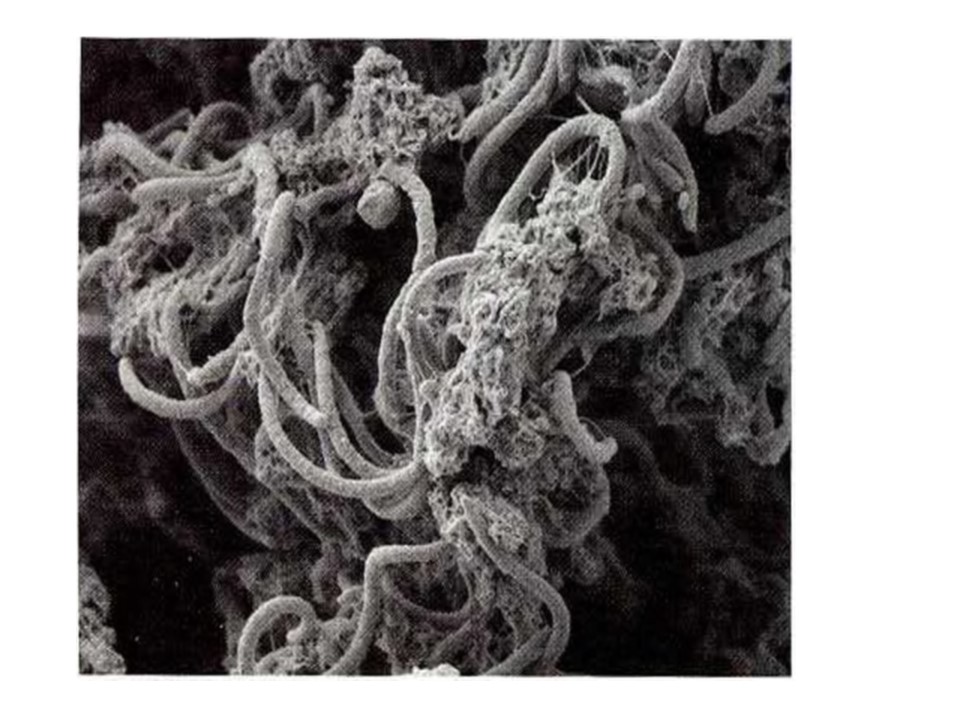
of the impact of biofilms in the food industry. A typical biofilm encrustation
is shown below:

Note the
significant growth of biofilm. In the reference by Pervical et al they have
extensive discussions regarding the development process in potable water. The
effect of chlorine in the water does diminish the growth slightly but it is a
common factor not only in loss of flow but in contamination. Fundamentally the
process is some three steps as shown below:


Note in the above we must have the layer irreversibly
adsorbed to allow an initial reversible bacterium to attach. We will explain
this later when examining the issues regarding forces. From Fleming and Ridgeway, we have:
The term “biofouling” is referred to as the undesired
development of microbial layers on surfaces. This operationally defined term
has been adapted from heat exchanger technology where “fouling” is defined
generally as the undesired deposition of material on surfaces, including:
– Scaling, mineral
fouling: deposition of inorganic material precipitating on a surface
– Organic fouling: deposition of organic substances (e.g.
oil, proteins, humic substances)
– Particle fouling: deposition of, e.g., silica, clay,
humic substances and other particles
– Biofouling: adhesion of microorganisms to surfaces and
biofilm development
The conditioning film shown above is essential. It is a
laying down of proteins and water which adhere to the surface via an adsorption
process. We generally suspect that such an adsorption is due to the van der
Walls forces from the surface to the structure of the specific proteins. We
will expand this discussion latter. Upon completion of the surfacing then the
proteins extending from the bacillus manage to penetrate this barrier and also
become attached via van der Waals forces. Darouiche discusses the impact of
biofilms on medical implants as well. He notes:
The essential factor in the evolution and persistence of
infection is the formation of biofilm around implanted devices. Soon after
insertion, a conditioning layer composed of host-derived adhesins (including
fibrinogen, fibronectin, and collagen) forms on the surface of the implant and
invites the adherence of free-floating (planktonic) organisms. Bacterial cell
division, recruitment of additional planktonic organisms, and secretion of
bacterial products (such as the glycocalyx) follow.
As Batte et al note:
Most of the pipes used in drinking water distribution
systems are made of plastic (PVC, PE, etc.) or metal (copper, cast iron) which
can become highly corroded (Figure 11). A recent survey of public distribution
system pipes in France showed that a large proportion of them are PVC (40%),
while the rest are grey iron (22%) or ductile iron (20%) (Cador 2002).
They continue:
The effects of the organic nutrients released by plastic
pipes on bacterial growth in drinking water have long been questioned. Organic additives
which leach out of plastic have a measurable impact on biofilm accumulation,
and are known to promote the multiplication of opportunistic, pathogenic
bacteria in laboratory tests. However, no field studies have looked at these
events…The lack of information on biofilm dynamics is a limiting
factor in managing the quality of water in distribution system and conducting
drinking water surveys. In spite of the difficulty of gaining access to the
inner surfaces of distribution pipes, biofilm measurement on pipe walls is
indispensable if more information on the water contamination risks is to be
obtained. New methods need to be developed, adapted, evaluated and optimized.
Such methods will create important advantages: continuous, non-destructive, simple,
in situ, online information on biofilm location and development.
From Preedy et al we have:
Biofilms are defined as a layer or layers of cells
adhered to a substratum which are generally embedded in an organic biological
matrix, i.e., extracellular polymeric substances (EPS). It is due to biofilm
formation that many bacteria survive in highly diverse and adverse environments
as a result of the polymicrobial ecosystem....Not surprisingly, biofilms have formed on a variety of
surfaces and are not only restricted to attachment at a solid—liquid interface
but have been observed at solid—air and liquid—liquid interfaces, with some
having beneficial results as well as detrimental; for example, in industry
biofilms are used successfully to separate coal particles from mineral matter.
On the other hand, biofilms have been known to cause biofouling reducing mass
and heat transfer and effectively increasing corrosion; also from a medical
point of view, biofilm colonized implanted medical devices often lead to
implant failure.
Current technological areas focus on several areas. The
areas are:
Nano Surface Enhancements: These are nanotechnology enhanced
titanium surfaces which demonstrate reduction in bacterial infection potential
and also demonstrate enhanced tissue and bone growth ensuring improved human
acceptance.
Surface Bactericidals for Intracorporeal Applications: These
are nanotechnologies for surface coatings of various catheters and the like
that result in dramatically reduced risks of infection by inhibiting bacterial
growth.
Selenium Enhanced Bactericidals: This is a selenium based
product which enables the control of bacterial growth. It appears to function
as a bacteriostatic agent. Combined with a bactericidal agent the combination
may affect dramatic control for long periods of bacteria on surfaces. This area
of product development appears to have several areas of application: (1) Those
applications which can be seen to be applied directly to the skin (cosmetics,
wound dressing, etc.), and (2) Those applications which can be used in clinical
and consumer applications to treat surfaces for anti-bacterial purposes, (3)
The control of growth on various surfaces of harmful flora or fauna.
Treatments have developed an approach to mitigate the growth
of biofilm. This is via the treatment of the surface by nano processing. The
treatments may be either by addition of materials such as nano Se or by the
selective deletion of surfaces to create a similar nano surfacing effect by the
use of lipase and other similar surface treatments. For example, nano Se has
been demonstrated to slow down S. aureus proliferation at a dose-dependent rate.
Increased lag time (in 40 and 20 μg/mL doses) would allow for the body’s immune
system to attack bacteria before exponential growth. We demonstrate some of
these results below:

In the above we note that the regrowth of bacteria is
dramatically reduced by the application of a Se surface at concentrations of
20-40 micrograms/milliliter. It has also been observed that surface coatings of
a density of 100 ng/sq cm of 20-70 nm diameter nano Se on the surface are also
adequate. The question we pose herein is; what is the physical process that
causes this to occur? Bacterial efficacy has been demonstrated as shown below:
·
Gram positive
Staphylococcus epidermidis was decreased by several logs on SeNP-coated paper
towels
·
SeNP coatings have also
reduced gram negative Pseudomonas aeruginosa, E. coli, MRSA, and ampicillin
resistant E. coli

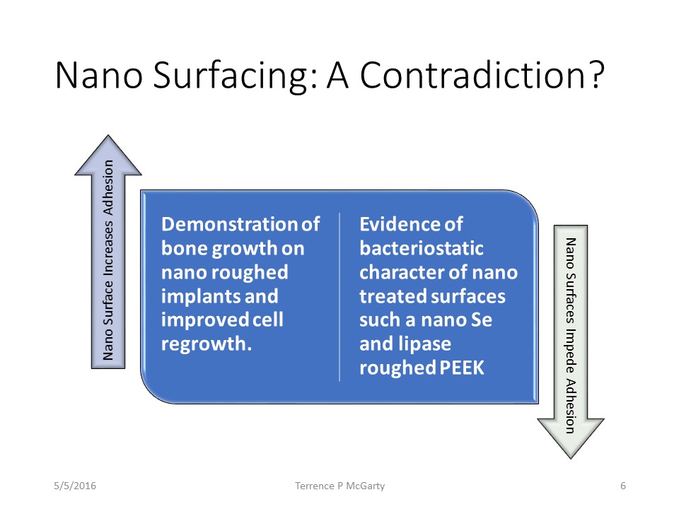
We now examine the physical processes which may account for
this twofold process. Namely:
1. Nano Se coatings and lipase nano processing of surfaces
tend to create a bacteriostatic environment.
2. Nano Se coatings and nano surface processing tend to
create an environment that enhances tissue adhesion.
These appear to be contradictory results. It would appear
that both processes are controlled by the same physical mechanism. Yet the
outcomes are dramatically different. We attempt to explain some of these
effects. However, it should be noted that in our analysis the explanation is
yet far from clear.
The following is a brief discussion of some of the basic
principles and specific technologies. Details are contained in the papers by
Webster and his team at Northeastern and Brown University. We have also
examined the literature in general and provides a summary regarding that as
well.
The principal basis for the technology is understanding
surface energy as relates to bacterial adhesion and subsequent biofilm growth.
We demonstrate the basic principle below for a eukaryotic cell using the
approach by Webster. On a smooth surface we have with cells a fibronectin, a
glycoprotein, which binds integrins. This allows the pathogen cell to attach to
the surface and commence biofilm growth.

The issue then is to create a surface which is not conducive
to the binding. This can be accomplished by manipulating the surface energy by
mechanical means. We can show that the protein absorption is proportional to
the surface energy. We can briefly examine van der Waals forces as discussed by
Butt and Kappl. Let us first consider simple Coulomb forces. We can consider
three types of surface to external adhesion for vdW. They are shown below:

Later we shall see that many argue for the simple connection
of inverse square where one surface is positive and the other is negative. This
is a simple vdW approach. However, the other two can be equally valid depending
on the nature of the molecules connecting. Namely in the case of proteins the
protein structure can be quite complex depending on the specific amino acid
construction. Note that for proteins the bonds generally are inverse fourth
power strength due to the dipole-monopole configuration. There may even be
cases in certain protein structures where the bonds are inverse sixth power[3].
The adhesion of bacteria to surfaces is a complicated and
yet to be satisfactorily answered phenomenon. There are several theories and we
will examine one herein. We use the DLVO approach which is a force or energy
approach. Alternative approaches using thermodynamically defined terms and
Gibbs Free Energy, G, have also been proposed but they do not seem to provide
adequate answers. Let us first review some general principles.
The DLVO (Derjaguin, Landau, Vervey, Overbeek) approach uses
the two forces; van der Waals and Ionic. The paper by Trefaly and Borkovec is
an excellent summary of this and we shall follow its approach.
Now the surface may be seen as below with these two forces:

Note that in close we have an attraction due to van der
Waals and then at a distance we have the double layer effect. The scales are
not precise but just descriptive. The vdW force is much stronger but there is a
positive "barrier" between it and the outer layer. Brownian motion
can get a bacterium close to the surface and catch it reversibly in the ionic
or DL area. However, to have an irreversible bond something must get to the vdW
section, a much stronger section.
Now the bacterium sends out a filament to try to bond to the
surface via the vdW forces. It must penetrate the barrier and then bond. In the
Boland et al paper we have an example of such bonding showing the extending
filaments:

They discuss what they term cellulose binding domains, CBD,
areas of the protein which do the binding in this case to cellulose. They
state:
These CBDs have been classified into 10 families (I-X) on
the basis of amino acid sequence homology. The amino acid sequences of CBDs in
C. cellulovorans and C. josui show high homology with those from other
cellulolytic genera such as Bacillus. CBDs in this family contain several
highly conserved amino acid sequences:
1.
Tryptophane-asparate-phenylalanine-asparagine-asparate-glycine-threonine
2. Isoleucine-alanine-alanine-isoleucine-proline-glutamine
3. Isoleucine-leucine-phenylalanine-valine-glycine
We can then ask; what if we roughen the surface, what will
that do? The specific answer is not known and even less understood
conceptually. A logical conclusion is that by roughing the surface we increase
the positive side by moving the inner vdW in and out and thus make it more
difficult to adhere. The Thermodynamic argument is a hand waving discussion of
surface energy. But we tried that argument above without success on adhesion of
human tissue cells.
As Bok noted in his Thesis:
The forces that govern microbial deposition, adhesion and
detachment are still not fully understood, and difficult to relate with each
other. In a previous study we successfully investigated the characteristic
shear force to prevent adhesion of microbial strains. In the current research
we used a more systematic approach by including not only the shear forces to
prevent adhesion, but also those that stimulate detachment of adhering
bacteria, as well as theoretical adhesion forces calculated using the extended
DLVO theory. …
1) A strong hydrodynamic shear force to prevent
adhesion relates to a strong hydrodynamic shear force to detach an adhering
organism. …
2) A weak hydrodynamic shear force to detach adhering
bacteria implies that more bacteria will be stimulated to detach by a passing
air-liquid interface through the flow chamber….
3) DLVO interactions determine the characteristic
hydrodynamic shear forces to prevent adhesion and to detach adhering
micro-organisms as well as the detachment induced by a passing air-liquid
interface. …
Thus from the above experimental analyses the DLVO has some
merit but it clearly does not describe the entire process. There are
significant issues still outstanding to be explained theoretically. Bacterial
adhesion and the formation of biofilms is still in the process of being fully
understood. Kanematsu and Barry provide an exceptionally strong discussion here
but we must resort to experimental data for phenomenological insight. Boland et
al also provide a substantial discussion on this but fail to provide a strong
analytical basis. Their analysis is useful to better understand some of the
phenomenology.
The thermodynamic paradigm is based upon certain principles
that aggregate large collections of common particles like gas, steam, or a
fluid. Thermodynamic principles work in the large like those used in reactors
or distillation columns or heat exchangers. We shall review some of these
principles and then demonstrate their lack of efficacy in this model.
For example, when considering the process of wetting, one
can generally use thermodynamic and surface tension methods. There is a
homogeneity on the surface and on the wetting materials. Tran and Webster
(2013) have provided an interesting analysis for nano scale wetting. They
explain it via the Wenzel and also the Cassie-Baxter models. They all involve
surface tension as is done in the core Young's analysis[4].
van Loosdrecht et al were one of the first to explain the
adhesion via thermodynamic principles. Then they state:
The Concept of Short-Range Interactions
If adhesion is performed at constant pressure and
temperature, and if the molecular composition of the surface does not change,
all G's can be replaced by the corresponding interfacial tensions. This concept
is restricted to those cases where bacteria and the solid surface are in direct
contact and the original phase boundaries are replaced by a new one, namely,
the bacterium solid interface. When this new interface is formed, interfacial
tensions may be used for a direct estimation of the adhesion Gibbs energy. …
The Concept of Long-Range Interactions
The DLVO theory for colloidal stability can be used to
calculate the interaction Gibbs energy between a particle and a surface as a
function of the separation distance (H). The balance of interracial Gibbs
energies … is the basic premise of this theory. The net interaction Gibbs
energy is interpreted in terms of Van der Waals interactions (which are usually
attractive) and an electric interaction due to the overlap of the electrical
double layers at the charged surfaces. The most important parameters
determining the van der Waals interaction are the Hamaker constant, which is a
material property, the distance (H) between bacterium and substratum, and the
geometry of the system. …
alAbas et al demonstrate oil pipeline biofilm as below:
Now in contrast alAbas et al note:
Thermodynamic approach
The thermodynamic approach assumes the system is in
equilibrium and the bacterial attachment is a reversible process. The
interfacial free energies between the interacting surfaces are compared and
calculated, …. This comparison is expressed in the so-called free energy of
adhesion. …The microbial adhesion will be favorable when the change in G, is
negative (< 0) and will not be energetically favorable if … positive.
They then continue:
DLVO Approach: The drawback of the thermodynamics
approach is that it ignores the electrical double-layer interaction with the
bacteria… This assumption is invalid as the bacterial cells have a
surface-negative or-positive charge. In contrast, the DLVO approach displays a
balance between attractive Lifshitz- van der Waals… and repulsive or attractive
electrostatic forces … These two forces are function of the distance (d)
between the bacteria and surface. In order to calculate the adhesion free
energy … the electrostatic interactions between surfaces should be included.
The inclusion of electrostatic interactions requires that the zeta potentials
of the interacting surfaces be measured, in addition to measuring contact
angles...Extended DLVO approach: The extended DLVO theory relates
the origin of hydrophobic interactions in microbial adhesion and includes four
fundamental interaction energies: Lifshitz-van der Waals, electrostatic, Lewis
acid-base, and Brownian motion forces …
The above approach
makes semi-macro thermodynamic assumptions. Specifically, a large mass of
surface, liquid and biofilm concentrate. In fact, the dynamics of the process
are totally overlooked. This is the general failing of thermodynamic approach;
they assume some form of steady state along with homogeneity. In reality we
have a dynamic process in a highly heterogeneous environment. We briefly discuss the technology to be employed. The
details are contained in the references by Webster discussed herein.
Nano surface treatments can be accomplished by treating the
surface itself or adding nano materials to the surface. The result is a stable
nano surface that inhibits bacterial growth and ensuing biofilm development. The
Gecko has nano fibrils on its feet that allow it to climb any surface by means
of van der Waals attraction as we see in nano material surfaces. The production
of nano Se is performed via a proprietary process but fundamentally is the
following:
Glutathione +NaOH + Se --à Nano Se
As Mendonca et al note, using reference to Webster's work,
the details of surface energy effects and adhesion or lack thereof:
The changes in initial protein–surface interaction are
believed to control osteoblast adhesion. This is a critical aspect of the
osseointegration process. When implants come into contact with a biological
environment, protein adsorption (e.g. plasma fibronectin) that occurs
immediately will mediate subsequent cell attachment and proliferation. Cell
binding to protein domains of adhesive extracellular matrix proteins involves
receptors termed integrin receptors that transmit signals through a collection
of proteins on the cytoplasmic face of the contact, termed focal contacts. … Webster and colleagues observed an increased
vitronectin adsorption on nanostructured surfaces when compared to conventional
surfaces. They also found an increased osteoblast adhesion when compared to
other cell types, such as fibroblasts, on the nanosurfaces. … Surface roughness
at the nanoscale is an important determinant of protein interactions that
ultimately direct cell activity in control of tissue formation at implant
surfaces.
To obtain a proper nano surface there are two methods. The
additive method uses nano Se which can be made at specific nano size and in a
very well controlled and defined distribution so as to assure the proper
surface energy. The second approach is the deletive approach whereby a nano
surfacing has a process that removes materials in such a controlled manner so
as to achieve the same desired surface energy.
Nano Selenium has been demonstrated as highly effective. In
addition, as we demonstrate below it is also safe and sustains the effect on
the surface for an extended period of time.
Why Se? The reasons are as follows:
- Essential micronutrient metalloid, and component of several key antioxidants, detoxifying and metabolic enzymes, in form of selenocysteine, selenomethionine
- Two allotropes: red (bioactive) and grey (crystalline) and Strong associations with reduction of Reactive Oxygen Species1,2,3 (ROS) as well as Cofactor of glutathione peroxidase
- Antibacterial activity to a broad range of pathogenic strains
- Nano Se can be produced at specific nano diameters with minimal dispersion, Spherical in shape
- Monodisperse—size distribution fits within one bell curve and negatively charged (uncoated)
The deletive approach used extraction mechanisms which
produce similar effects to the additive mechanism of nano Se. The advantage of
such an approach is that it does not add anything to the material. The
disadvantage in certain active biological surfaces such as human skin is that
it causes immunological effects. However, its used in stable media such as
PEEK, Titanium, steel, and other materials used for water flow and containment
is that it can be readily effected and at low cost. There are numerous
processes to implement nanoscale surface features on metallic or polymeric
surfaces. We then utilize one of our processes to create such nanoscale features:
Anodization or Chemical etching. The deletive approach provides comparable
results to that for Se coatings.
There have been a variety of biofilm inhibition methods. As
Garrett et al note:
Bacterial adhesion has become a significant problem in
industry and in the domicile, and much research has been done for deeper
understanding of the processes involved. A generic biological model of
bacterial adhesion and population growth called the bacterial biofilm growth
cycle, has been described and modified many times.
The biofilm growth cycle encompasses bacterial adhesion
at all levels, starting with the initial physical attraction of bacteria to a
substrate, and ending with the eventual liberation of cell clusters from the biofilm
matrix. When describing bacterial adhesion one is simply describing one or more
stages of biofilm development, neglecting the fact that the population may not
reach maturity. This article provides an overview of bacterial adhesion, cites
examples of how bacterial adhesion affects industry and summarizes methods and
instrumentation used to improve our understanding of the adhesive properties of
bacteria.
The NRC report states[5]:
The pipe surface itself can influence the composition and
activity of biofilm populations. Studies have shown that biofilms developed
more quickly on iron pipe surfaces than on plastic PVC pipes, despite the fact
that adequate corrosion control was applied, the water was biologically treated
to reduce AOC levels, and chlorine residuals were consistently maintained…In
addition to influencing the development of biofilms, the pipe surface has also
been shown to affect the
composition of the microbial communities presents in the
biofilm. Iron pipes supported a more diverse microbial population than did PVC
pipes. The purpose of these studies is not to indicate that certain pipe
materials are preferred over another but to demonstrate the importance of
considering the type of materials that come into contact with potable water.
We examined several issues. Specifically:
1. What is a Biofilm? This we have answered by reference to
various studies.
2. How do biofilms form? The answer to this may often depend
but it is clearly a dynamic process.
3. What is the physical phenomenon that allows biofilms to
adhere and have strong adsorption? This is a work in progress. We believe the
thermodynamic approach is problematic at best. It is necessary to consider more
detailed dynamic physical phenomenon. We make some suggestions here.
4. What is the effect of nano-surfacing on biofilms? This
appears to be uncertain at best. There are contrasting phenomenological
results.
5. Why does nano-surfacing enhance adsorption of certain
eukaryotic cells such as bone and ligaments while inhibiting the adsorption of
prokaryotic cells such as bacteria? This appears not to have been examined.
6. How can nano-surfacing be optimized to minimize biofilms?
Argument from surface energy have been proposed but are problematic.
These questions can and have been answered in part but there
remains a set of uncertainties that challenge the effective utilization of nano
technologies.
We can possibly argue the following explanation from what we
have developed herein.
1. The first coating of a surface is by the protein layer.
Generally, this is done by some local van der Waals forces since the proteins
are close to the surface and are well known to exhibit such forces. Also the
protein layer seems to be a prerequisite for adhesion. However, the type of
protein layer may very well depend on the surface structure. They structure of
proteins vary widely and perhaps if we adjust the nanostructure we selectively
change the type of protein adhering to the surface.
2. We know that bacteria seem phenomenologically to require
proteins to adhere for them in turn to reversibly adhere to the proteins. This
the proteins must be electrostatically and vdW wise strongly attracted to the
surface and the cell.
3. After a reversible adhesion then we seem to have the
appearance of protein filaments extruding from the bacteria and down through
the protein layer, most likely using the protein to overcome the barrier wall
normally between van der Waals and electronic forces. Once the filament hits
the surface then it adheres irreversibly and the biofilm commences growth.
4. The supposition is that by changing the roughness of the
surface we change the types of proteins or the nature of their adhesions on the
surface. There does not appear to be any research determining this one way or
the other at this time.
The challenge of this analysis is shown below. On the one hand certain biologicals adhere on roughness and others are repelled. One of the continuing questions is why, and what is the fundamental physical reason.
1.
alAbbas, F., et al,
Bacterial attachment to metal substrate and its effects on
microbiologically-influenced corrosion in transporting hydrocarbon pipelines,
Paper Best Practices in Pipeline Operations Conf, Bahrain, 2012.
2.
Barghouti, S., A Universal
Method for the Identification of Bacteria Based on General PCR Primers, Indian
J Microbiol (Oct–Dec 2011) 51(4):430–444
3.
Barron, E., Rapid
Identification of Bacteria and Yeast: Summary of a National Committee for
Clinical Laboratory Standards Proposed Guideline, MEDICAL MICROBIOLOGY • CID
2001:33 (15 July) 221
4.
Batte, M., et al, Biofilms
in drinking water distribution systems, Reviews in Environmental Science and
Bio/Technology · January 2003
5.
Boe-Hansen, R.,
Albrechtsen, H.-J., Arvin, E. and Jørgensen, C. (2002a). Dynamics of biofilm
formation in a model drinking water distribution system. J. Water Supply Res.
Technol–Aqua., 51, 399–406.
6.
Boe-Hansen, R.,
Albrechtsen, H.-J., Arvin, E. and Jørgensen, C. (2002b). Bulk water phase and
biofilm growth in drinking water at low nutrient conditions. Wat. Res., 36,
4477–4486.
7.
Boe-Hansen, R.,
Albrechtsen, H.-J., Arvin, E. and Jørgensen, C. (2002c). Substrate turnover at
low carbon concentrations in a model drinking water distribution system. Wat.
Sci. Tech.: Water Supply, 2(4), 89–96.
8.
Boe-Hansen, R., et al,
Monitoring biofilm formation and activity in drinking water distribution
networks under oligotrophic conditions, Water Sci & Tech, Vol 47 No 5,
2003.
9.
Boks, N., Bacterial
interaction forces in adhesion dynamics, PhD Thesis, Groningen, 2009.
10. Boland, T., et al, Molecular Basis of Bacterial Adhesion,
Handbook of Bacterial Adhesion: Principles, Methods, and Applications, Edited
by: Y. H. An and R. J. Friedman, Humana Press Inc., Totowa, NJ
11. Butt H et al, Physics and Chemistry of Interfaces, Wiley (New
York) 2013.
12. Butt, H., M. Kappl, Surface and Interfacial Forces, Wiley (New
York) 2010.
13. Combs Jr, Gerald F., and William P. Gray. "Chemopreventive
agents: selenium." Pharmacology & therapeutics 79.3 (1998).
14. Costerton, J., et al, BACTERIAL BIOFILMS IN NATURE AND DISEASE, Ann.
Rev. Microbiol. 1987. 41:435
15. Darouiche, R., Treatment of Infections Associated with Surgical
Implants, NEJM 2004, 350;14
16. Davis, M., Water and Wastewater Engineering, McGraw Hill (New
York) 2010.
17. Elimelech, M., W. Phillip, The Future of Seawater Desalination:
Energy, Technology, and the Environment, Science, Vol 333, Aug 2011.
18. Emerson, D., et al, Identifying and Characterizing Bacteria in
an Era of Genomics and Proteomics, Bioscience, November 2008 / Vol. 58 No.
10
19. EPA, Control of Biofilm Growth in Drinking Water Distribution
Systems, EPA/625/R-92/001, Environmental Protection Development June 1992
20. Fleming, H., H. Ridgeway, Biofilm Control: Conventional and
Alternative Approaches, Biofilms (Springer) 2008
21. Garrett, T., et al, Bacterial adhesion and biofilms on surfaces,
Progress in Natural Science 18 (2008) 1049–1056
22. Gitai, Z., The New Bacterial Cell Biology: Moving Parts and
Subcellular Architecture, Cell, Vol. 120, 577–586, March 11, 2005
23. Gorth, D., S. Puckett, B. Ercan*, T.J. Webster, “Decreased
bacteria activity on Si3N4 surfaces compared with PEEK or titanium,”
International Journal of Nanomedicine, 7: 4829-4840 (2012).
24. Gupta, R., Hydrology and Hydraulic Systems, Waveland (Long Grove
IL) 2008
25. Huang, T., et al, Composite Surface for Blocking Bacterial
Adsorption on Protein Biochips, BIOTECHNOLOGY AND BIOENGINEERING, VOL. 81, NO. 5,
MARCH 5, 2003
26. Irvine, D., et al, Simulations of Cell-Surface Integrin Binding
to Nanoscale-Clustered Adhesion Ligands, Biophysical Journal Volume 82 January
2002 120–132
27. Kanematsu, H., D. Barry, Biofil and Materials Science, Springer
(New York) 2015
28. Katsikogianni, M., Y. Missirilis, Concise Review of Mechanisms
of Bacterial Adhesion to Biomaterials and of Techniques Used in Estimating
Bacterial Material Interactions, Euro Cells and Materials, Vol 8 2004
29. Khang, D., S.Y. Kim, P. Liu-Synder, G.T.R. Palmore, S.M. Durbin,
T.J. Webster, “Enhanced fibronectin adsorption on carbon
nanotubes/poly(carbonate) urethane: independent role of surface nano roughness
and associated surface energy,” Biomaterials, 28(32):4745-4768 (2007).
30. Krasowska, A., K. Sigler, How microorganisms use hydrophobicity
and what does this mean for human needs? Frontiersin Cellular and Infection Microbiology,
August 2014, Volume4, Article112
31. Kulkarni, S., Nanotechnology: Principles and Practices, Springer
(New York) 2011
32. Kumar et al, “Selenium nanoparticles involve HSP-70 and SIRT1 in
preventing the progression of type 1 diabetic nephropathy”, Chemico-Biological
Interactions 223 (2014).
33. Machado, M., et al, Decreased Staphylococcus aureus biofilm
formation on nanomodified endotracheal tubes: a dynamic airway model, International
Journal of Nanomedicine 2012:7 3741–3750
34. McFaddin, J., Biochemical Tests for the Identification of
Medical Bacteria, William and Wilkens, (Baltimore) 1980.
35. Mendonca, G., et al, Advancing dental implant surface technology
– From micron to nano topography, Biomaterials 29 (2008) 3822–3835
36. National Research Council, Identifying Future Drinking Water
Contaminants, 1998, http://www.nap.edu/catalog/9595.html
37. Percival, S., et al, Microbiological Aspects of Biofilms and
Drinking Water, CRC (New York) 2000.
38. Perla, V., T. Webster, Better osteoblast adhesion on
nanoparticulate selenium— A promising orthopedic implant material, Published
online 29 July 2005 in Wiley InterScience (www.interscience.wiley.com ). DOI:
10.1002/jbm.a.30423
39. Preedy, E., et al, Surface Roughness Mediated Adhesion Forces
between Borosilicate Glass and Gram-Positive Bacteria, www.pubs.acs.org/Langmuir Langmuir
2014, 30, 9466—9476
40. Ramos, J., T.J. Webster, “Cytotoxicity of selenium nanoparticles
in rat dermal fibroblasts,” International Journal of Nanomedicine, 7: 3907-3914
(2012).
41. Schkolnik, S., et al, In Situ Analysis of a Silver
Nanoparticle-Precipitating Shewanella Biofilm by Surface Enhanced Confocal
Raman Microscopy, PLOS One December 28, 2015
42. Srey, S., et al, Biofilm formation in food industries: A food
safety concern, Food Control 31 (2013) 572e585
43. Srivastava et al, In vivo synthesis of selenium nanoparticles by
Halococcus salifodinae BK18 and their anti-proliferative properties against
HeLa cell line. Biotechnology Progress (2014)
44. Steel, E., Water Supply and Sewerage, McGraw Hill (New York)
1953.
45. Tran, P, T.J. Webster, Antimicrobial selenium nanoparticle
coatings on polymeric medical devices, Nanotechnology, 24 (15): 155101 (2013).
46. Tran, P. and T.J. Webster, Selenium nanoparticles inhibit
Staphylococcus aureus growth, International Journal of Nanomedicine,
6:2001-2011 (2011).
47. Tran, P., and T.J. Webster, “Enhanced osteoblast adhesion on
nanostructured selenium compacts for anti-cancer orthopedic applications,”
International Journal of Nanomedicine, 3(3): 238-247 (2008).
48. Tran, P., L. Sarin, R. Hurt, and T.J. Webster, Titanium surfaces
with adherent selenium nanoclusters as a novel anti-cancer orthopedic material,
Journal of Biomedical Materials Research Part A, 93A (4): 1417-1428 (2010).
49. Tran, P., T.J. Webster, Understanding the wetting properties of
nanostructured selenium coatings: the role of nanostructured surface roughness
and air-pocket formation, International Journal of Nanomedicine, 8: 2001-2009
(2013).
50. Trefalt, G., M. Borkovec, Overview of DLVO Theory, www.colloid.ch/dlvo (2014)
51. U.S. Fish and Wildlife Service, 3.8 Bacterial Identification
Techniques, https://www.google.com/url?sa=t&rct=j&q=&esrc=s&source=web&cd=5&ved=0ahUKEwj4-9HFrqfMAhUCMj4KHejVD3UQFghPMAQ&url=http%3A%2F%2Fwww.fws.gov%2Fpacific%2Ffisheries%2Ffishhealth%2Fdocuments%2Fbluebook%2Fcdr_pdfs%2Findexed%2Fb3_08.pdf&usg=AFQjCNGbL5FE3e0woiPb-DfOk-xEKRBKqA&sig2=aszx3gob3awPK32KVVj35A&cad=rjt
52. Van der Kooij, Potential for biofilm development in drinking
water distribution systems, Journal of Applied Microbiology Symposium
Supplement 1999,85, 39-4s
53. van Loosdrecht, M, et al, Bacterial Adhesion: A Physiochemical
Approach, Microb Ecol 1989 V 17
54. Voutchkov, N., Desalination Engineering, McGraw Hill (New York)
2013.
55. Wang, Q., T. Webster, Nanostructured selenium for preventing
biofilm formation on polycarbonate medical devices, Published online 15 June
2012 in Wiley Online Library (wileyonlinelibrary.com). DOI: 10.1002/jbm.a.34262
56. Wang, Q., T.J. Webster, Short communication: Inhibiting biofilm
formation on paper towels through the use of selenium nanoparticles coatings,
International Journal of Nanomedicine, 8: 407-411 (2013).
57. Webster, T, A.A. Patel, M.N. Rahaman, Anti-infective and
osteointegration properties of silicon nitride, poly-ether-ether-ketone, and
titanium implants (vol 8, pg 4447, 2012),” Acta Biomaterialia, 10 (3):
1485-1486 (2014).
58. Werber, J., et al, Materials for next-generation desalination
and water purification membranes, NATURE REVIEWS MATERIALS, 2016
59. Xu, X., D., Mosher, Fibronectin and Other Adhesive Glycoproteins,
R.P. Mecham (ed.), The Extracellular Matrix: an Overview, Biology of
Extracellular Matrix, DOI 10.1007/978-3-642-16555-9_2, # Springer-Verlag Berlin
Heidelberg 2011
60. Zhang and Gladyshev, “General Trends in Trace Element
Utilization Revealed by Comparative Genomic Analyses of Co, Cu, Mo, Ni and Se”,
J. of Biol. Chem (2010).


