Tuesday, July 31, 2012

Recession Statistics Q2 2012

We have examined and comment here on the St Louis Fed's Recession Statistics. We have been tracking them quarterly since the beginning of the collapse and frankly they are appearing to get worse. Given what we see and other data we have reported upon there is a strong possibility of a second recession this Fall.

Let us examine the data.
Industrial Production appears on average, weak but on par. This frankly is the best metric.
Income is below the lowest. Income is just not recovering and in fact it will be the driver for the next dip. Despite Production, if there are no customers then we will see that drop also.
Employment is very weak. Not the worst yet but getting there. It shows no sign of any improvement and this will drag on well into 2014.
Retail Sales seems to be keeping up but I suspect that a good deal of that is credit increase as well as Government Supports; Unemployment, FICA elimination and Food Stamps. These Government Programs are non job creating and just add drag to the economy.

The GDP stats show we are now below the lowest. This is truly a concern. The weak growth rate will not see any improvement under the current administration.
Personal Consumption, an element of the GDP, is well below the lowest. People are just not buying, not enough income and employment.
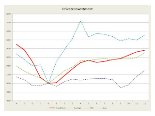
However Private Investment is above average. Money is around but the investments do not result in employment, just improvements in productivity.
Government Consumption is the lowest yet, and much of this is State and Local.
Exports are high relative...
Imports are average.

The above presents a dire forecast for 2013-2014, especially if we remain on course.

Monday, July 30, 2012

Melanoma, Sun Damage and Pathways


There is extensive epidemiological data indicating that melanoma is caused by UV radiation. Now there has been, up to this date, little information relating specific effects of UV radiation to specific causative gene changes. In a recent article in Cell by Hodis etal, the authors relate the impact of sun damage on melanocytes and the initiation of melanoma[1].This is an interesting paper and the approach is quite innovative and worth examining.

The authors summarize their work as follows:

Despite recent insights into melanoma genetics, systematic surveys for driver mutations are challenged by an abundance of passenger mutations caused by carcinogenic UV light exposure.

We developed a permutation-based framework to address this challenge, employing mutation data from intronic sequences to control for passenger mutational load on a per gene basis.

Analysis of large-scale melanoma exome data by this approach discovered six novel melanoma genes (PPP6C, RAC1, SNX31, TACC1, STK19, and ARID2), three of which—RAC1, PPP6C, and STK19—harbored recurrent and potentially targetable mutations.

Integration with chromosomal copy number data contextualized the landscape of driver mutations, providing oncogenic insights in BRAF- and NRAS-driven melanoma as well as those without known NRAS/BRAF mutations.

The landscape also clarified a mutational basis for RB and p53 pathway deregulation in this malignancy. Finally, the spectrum of driver mutations provided unequivocal genomic evidence for a direct mutagenic role of UV light in melanoma pathogenesis.

In a release from MD Anderson Cancer Center they state[2]:

By creating a method to spot the drivers in a sea of passengers, scientists at the Broad Institute of MIT and Harvard, the Dana-Farber Cancer Institute and The University of Texas MD Anderson Cancer Center have identified six genes with driving mutations in melanoma, three of which have recurrent 'hotspot' mutations as a result of damage inflicted by UV light. Their findings are reported in the July 20 issue of the journal Cell.

"Those three mutations are the first 'smoking gun' genomic evidence directly linking damage from UV light to melanoma," said co-senior author Lynda Chin, M.D., Professor and Chair of MD Anderson's Department of Genomic Medicine. "Until now, that link has been based on epidemiological evidence and experimental data."

"This study also is exciting because many of the recent large-scale genomic studies have not discovered new cancer genes with recurrent hot-spot mutations, a pattern strongly indicative of biological importance," said Chin, who also is scientific director of MD Anderson's Institute for Applied Cancer Science.

The six new melanoma genes identified by the team are all significantly mutated and provide potential targets for new treatments.

Let us first detail several of these genes.

1. RAC1

From NCI we have RAC1 located at 7p22 and described as follows[3]:

The protein encoded by this gene is a GTPase which belongs to the RAS superfamily of small GTP-binding proteins. Members of this superfamily appear to regulate a diverse array of cellular events, including the control of cell growth, cytoskeletal reorganization, and the activation of protein kinases.

From the NCI Pathway database we have a complex set of pathway interactions[4]. In a similar manner we can examine the pathways from the MMMP data base[5]. In all cases of this gene and the others recenbctly elucidated, the pathways are partially informative and need additional investigation.

2. PPP6C

From NCI we have PPP6C located at 9q33.3 and described as follows[6]:

This gene encodes the catalytic subunit of protein phosphatase, a component of a signaling pathway regulating cell cycle progression. Splice variants encoding different protein isoforms exist.

3. STK19

From the NCI database this gene is located at 6q21.3 and functions as follows[7]:

This gene encodes a serine/threonine kinase which localizes predominantly to the nucleus. Its specific function is unknown; it is possible that phosphorylation of this protein is involved in transcriptional regulation. This gene localizes to the major histocompatibility complex (MHC) class III region on chromosome 6 and expresses two transcript variants

Thus the genes perform a broad and generally non-correlative set of functions. The authors have argued that the genes are targetable as say with BRAF but a more complete understanding of full pathway interactions would be essential.

Counting UV Hits

The authors discuss the fact that m UV mutations convert C (cytidine) to T (thymidine). Now as Watson et al have shown pp 204-209[8]) when cytidine is methylated as shown below the uridine product is converted to thymidine and this there is a mis-reading of the DNA. These C to T transitions are caused in the case of melanoma often by UV. We have also argued that they may equally be caused by backscatter X-rays which have enough energy to break bonds to cause a methylation as well. 


Now the principle the authors employed was a presumptive one based upon the following:

1. C to T transition is random across the DNA sequences.

2. The average number of such transitions should be the same in both Introns and Exons.

3. Exons express genes which do things.

4. If there are a statically larger number of C to T transitions in a specific Exon, i.e. a gene say Gene X, then that gene most likely is causative of the melanoma.

We demonstrate this concept in the diagram below:

 Now it was through a process of this type which allowed the authors to identify a collection of twelve genes, six known to be related to melanome, and six not previously known to be related, to be presumptively causitive of the malignancy.

From an article in Science Daily they state, using a somewhat less than precise metaphor, the following[9]:

By creating a method to spot the drivers in a sea of passengers, scientists at the Broad Institute of MIT and Harvard, the Dana-Farber Cancer Institute and The University of Texas MD Anderson Cancer Center have identified six genes with driving mutations in melanoma, three of which have recurrent 'hotspot' mutations as a result of damage inflicted by UV light. Their findings are reported in the July 20 issue of the journal Cell.

"Those three mutations are the first 'smoking gun' genomic evidence directly linking damage from UV light to melanoma," said co-senior author Lynda Chin, M.D., Professor and Chair of MD Anderson's Department of Genomic Medicine. "Until now, that link has been based on epidemiological evidence and experimental data."

"This study also is exciting because many of the recent large-scale genomic studies have not discovered new cancer genes with recurrent hot-spot mutations, a pattern strongly indicative of biological importance," said Chin, who also is scientific director of MD Anderson's Institute for Applied Cancer Science.

The six new melanoma genes identified by the team are all significantly mutated and provide potential targets for new treatments.

Puzzle has thousands of potential pieces, but only requires a few dozen. A number of important mutations had previously been identified as melanoma drivers. These include BRAF (V600) mutations, present in half of all melanomas, and NRAS (Q61) mutations. However, the vast majority of these mutations do not appear to be caused by direct damage from UV light exposure.

UV light causes many mutations of genes in melanocytes. The mutations occur in both introns and exons. The question is which of these mutations is significant and for example is there a level at which they become malignant. An interesting question can be asked about melanoma in situ, the early stage of melanoma where the melanocytes have enlarged nucleoli and express a loss of localization. It is well known histologically that MIS is often discovered in sun damaged areas. Thus one would suspect that at this early stage many of this methylation like changes doe to UV radiation is present.

The article continues:

To counter this effect, the researchers turned to parts of the genome that don't code for proteins, called introns, and other inactive DNA segments that flank exons. By comparing the frequency of mutations in the inactive segments to the frequency of mutations in the exons, the researchers built a framework for assessing the statistical significance of functional mutations.

Approach identifies six known cancer genes, six new ones.

The analysis identified functional mutations in the well-known cancer genes BRAF, NRAS, PTEN, TP53, CDKN2A and MAP2K1.

It also uncovered five new genes, RAC1, PPP6C, STK19, SNX31, and TACC1.

Most are associated with molecular pathways involved in cancer but had not been previously recognized as significantly mutated in melanoma. Their presence in the tumor samples ranged from 3 percent to 9 percent.

The sixth new gene tied to melanoma was ARID2, an apparent tumor-suppressor gene possessing a significant number of loss-of-function mutations found in 7% of patient samples.

"Six new melanoma genes have been picked out from thousands of mutated genes," said Eran Hodis, co-lead author who is a computational biologist in the Garraway lab at the Broad Institute and an M.D.-Ph.D. student at Harvard and MIT. "The same approach may bring clarity to genome sequencing studies of other cancers plagued by high passenger mutation rates, for example lung cancer." ...


Most exciting, three of the discovered genes possessed 'hotspot' mutations found in the exact same position in multiple patients providing another line of evidence indicating these mutations contribute to melanoma.

"We have now discovered the third most common hotspot mutation found in melanoma is present in a gene called RAC1, and unlike BRAF and NRAS mutations, this activating mutation is attributable solely to characteristic damage inflicted by sunlight exposure" said Ian R. Watson, Ph.D.,...

Observations

This is a significant contribution in my opinion. It also, in my opinion, raises some very interesting questions.

1. How many hits are required to make the change?

2. What are the pathway effects that result in malignancy?

3. How does MIS fit within this model?

4. If UV radiation can do this then we would expect that X rays would have equal effects and if so then backscatter X rays which penetrate just enough would be of significance. If that is correct how much radiation would be required?

5. If we have these putative genes and there are targets, then how easy would it be to develop anti-cancer drugs for these targets?

6. If we see BRAF failure and return of the malignancy then is it possibly from these new genes, if so which ones, and if some of them in what order of importance?

7. When performing biopsies on melanomas, should examination for these genes be a common practice?

This paper raises many more such questions.
 
References

Hodis, E., et al, A Landscape of Driver Mutations in Melanoma, Cell, Volume 150, Issue 2, 251-263, 20 July 2012.

Sunday, July 29, 2012

MDM4 and Melanoma: More Pathways


Recent efforts in managing melanoma have focused upon BRAF and a mutation of a V600 BRAF form. By targeting this mutation and the pathway related thereto one can find ways to block the mutated pathway and this in principle block the continuing metastasis. This works for a while and then the cells find ways around this. There are undoubtedly many other changes in cellular pathways that result in uncontrolled proliferation and failure of apoptosis. Namely the cells continue to grow and fail to die off.

Focus on other pathway defects is continuing and there has been recent focus on MDM4, which is a control element of p53, the product of TP53 which is a key control element of proliferation and apoptosis. In a recent paper by Gembarska et al the authors state the following[1]:

The inactivation of the p53 tumor suppressor pathway, which often occurs through mutations in TP53 (encoding tumor protein 53) is a common step in human cancer. However, in melanoma—a highly chemotherapy-resistant disease—TP53 mutations are rare, raising the possibility that this cancer uses alternative ways to overcome p53-mediated tumor suppression. Here we show that Mdm4 p53 binding protein homolog (MDM4), a negative regulator of p53, is upregulated in a substantial proportion (~65%) of stage I–IV human melanomas and that melanocyte-specific Mdm4 overexpression enhanced tumorigenesis in a mouse model of melanoma induced by the oncogene Nras.

MDM4 promotes the survival of human metastatic melanoma by antagonizing p53 proapoptotic function. Notably, inhibition of the MDM4-p53 interaction restored p53 function in melanoma cells, resulting in increased sensitivity to cytotoxic chemotherapy and to inhibitors of the BRAF (V600E) oncogene. Our results identify MDM4 as a key determinant of impaired p53 function in human melanoma and designate MDM4 as a promising target for antimelanoma combination therapy.

Now MDM4, also called Mdm4 p53 binding protein homolog, is located at 1q32. It acts in a somewhat complex manner to control p53 functions. From NCI we have the following description of the gene and its product[2]:

This gene encodes a nuclear protein that contains a p53 binding domain at the N-terminus and a RING finger domain at the C-terminus, and shows structural similarity to p53-binding protein MDM2. Both proteins bind the p53 tumor suppressor protein and inhibit its activity, and have been shown to be overexpressed in a variety of human cancers. However, unlike MDM2 which degrades p53, this protein inhibits p53 by binding its transcriptional activation domain. This protein also interacts with MDM2 protein via the RING finger domain, and inhibits the latter's degradation. So this protein can reverse MDM2-targeted degradation of p53, while maintaining suppression of p53 transactivation and apoptotic functions.

The sources for information on p53 pathway and its relation to MDM4 are extensive[3]. Specific details of the p53 pathway are shown in the NCI data[4] bases for pathways. However, we shall present a simplified description based upon KEEG pathway data. This we do below (We combine from the KEGG genome database[5]).


Note in the above we have a complex control path between MDM2 and MDM4 with p53. The p53 is activated when DNA damage is perceived or from other factors. p53 then activates a collection of pathways which in turn block the cell cycle or initiate apoptosis. If p53 does not function then we have an uncontrolled cell. The control of p53 can be blocked by MDM4 blockage as shown above. That is the principle which the authors have presented.

Prior work by Macchiarulo et al stated the following regarding this combination[6]:

Alterations of p53 signalling pathway is the most frequent event in human cancers. About 50% of these, albeit showing wild-type p53, have flaws in the control mechanisms of p53 levels and activity. MDM2 and MDMX (MDM4) are the main negative regulators of p53.

The relevance of MDM2 on the regulation of p53 levels and activity has fostered the development of strategies aimed at restoring p53 functions by blocking the physical interaction between MDM2 and p53. As a consequence, a number of different small molecules and peptidomimetics have been disclosed in the last decade as inhibitors of MDM2/p53 interaction.

Recent studies, however, have thrust MDMX into the limelight as an additional chemotherapeutic target, suggesting the presence of a more complex relationship between MDM2, MDMX and p53. In this review article, we report key aspects of MDMX-mediated regulation of p53, recent advances in the structural characterization of the protein, and the progress made so far in the medicinal chemistry of MDMX ligands.

Note that MDMX is now called MDM4, to avoid confusion. The Macchiarulo paper was published a year ago (2011) and it presented the connection of MDM4 and loss of p53 control in a broader context of cancer development and spread. The Gembarska paper on the other hand has focused on melanoma. Earlier work was performed in a Doctoral Thesis in 2007 in Rotterdam, by Meulmeester who states[7]:

The p53 tumor suppressor gene encodes a sequence-specific transcription factor whose activity is either disabled or attenuated in the vast majority of human cancers. Its inactivation occurs in about 50% of human tumors through mutations affecting the p53 locus directly.

p53 transcriptionally activates a vast, constantly growing number of target genes, resulting in various biological outcomes such as cell-cycle arrest and apoptosis. Several types of stress, such as oncogene activation, hypoxia and DNA damage, result in an increase in p53 levels and the subsequent activation of p53 target genes (Vogelstein et al., 2000). One of the best-characterized target genes of p53 is the mdm2 gene, which contains two promoters.

The first promoter (P1) drives mdm2 expression constitutively (Jones et al., 1996), while p53 binds two adjacent p53-responsive elements within the second promoter (P2), thereby promoting transcription of the mdm2 gene. Under normal circumstances, p53 is tightly regulated through the interaction with its negative regulator Mdm2, which counteracts p53 function in a number of ways.

 The autoregulatory negative feedback loop, whereby p53 induces Mdm2 expression resulting in the repression of p53 function, most probably serves as an important mechanism to restrain p53 activity in normal cells. Therefore, uncontrolled, high expression of Mdm2 may result in improper inactivation of p53 function. It has been shown that in 5-10% of all human tumors Mdm2 is overexpressed, due to gene amplification, transcriptional- or posttranscriptional mechanisms. In most of these cases the p53 gene is wild type, presumably because Mdm2 overexpression alleviates the selective pressure for direct mutational inactivation of the p53 gene.

As regards to the pathway discussion we presented above Meulmeester remarks:

The complex web of ATM-mediated activation of the p53 pathway. ATM mediates direct and indirect phosphorylation of p53, while 14-3-3 binding to p53 is augmented by ATM-mediated de-phosphorylation of p53. Phosphorylation of Strap by ATM results in the recruitment of Strap/p300 complexes towards p53 that elevates its acetylation.

A safeguard mechanism exists to ensure proper p53 activation by inhibiting its inhibitors Mdm2 and Mdmx. Phosphorylation of Mdmx/Mdm2 attenuates their interaction with the ubiquitin protease HAUSP, resulting in the instability of Mdmx and Mdm2. Thus ATM activates p53 via a sophisticated mechanism, while it ensures proper activation by inhibition of its negative regulators .

Again note that MDMX is now MDM4. He does raise the issue of a complex feedback loop which may have some internal instabilities. Namely the loop between MDM2, MDM4, and p53 may have unstable points under certain conditions. Some have approached this via rate reaction equations but as we have discussed elsewhere the rate reaction equations require large concentrations. In a cell we have a few protein molecules with limited binding sites. This specific low density case has particular concerns of instabilities. Thus there may not just be a mutation of MDM4 but also some instabilities in the internal dynamics.

In a paper by Mancini et al (2009), the authors state[8]:

MDM4 is a key regulator of p53, whose biological activities depend on both transcriptional activity and transcription independent mitochondrial functions. MDM4 binds to p53 and blocks its transcriptional activity; however, the main cytoplasmic localization of MDM4 might also imply a regulation of p53-mitochondrial function.

Here, we show that MDM4 stably localizes at the mitochondria, in which it (i) binds BCL2, (ii) facilitates mitochondrial localization of p53 phosphorylated at Ser46 (p53Ser46P) and (iii) promotes binding between p53Ser46P and BCL2, release of cytochrome C and apoptosis. In agreement with these observations, MDM4 reduction by RNA interference increases resistance to DNA-damage-induced apoptosis in a p53-dependent manner and independently of transcription.

Consistent with these findings, a significant downregulation of MDM4 expression associates with cisplatin resistance in human ovarian cancers, and MDM4 modulation affects cisplatin sensitivity of ovarian cancer cells. These data define a new localization and function of MDM4 that, by acting as a docking site for p53Ser46P to BCL2, facilitates the p53-mediated intrinsic-apoptotic pathway. Overall, our results point to MDM4 as a double-faced regulator of p53.

Thus they make the connection of control of MDM4 products as key to the ultimate functionality of p53. They also have examined the effects of cisplatin in the case of ovarian cancers. They did not examine ways to block MDM4 and its protein. The principle here is that the pathway control element such as p53 can be dysregulated by another gene MDM4. The question of course is what has happened to MDM4. We address that later.

In a discussion of the paper by Gembarska et al, Azvolinsky then states[9]:

While the TP53 gene, which encodes the tumor protein 53, is found mutated in the vast majority of tumors, TP53 is intact in more than 95% of melanomas. The p53 tumor suppressor pathway is important for most cancers, preventing neoplastic growth, and inactivation of the pathway is a common driver mutation for cancer.

Exploring alternative mechanisms of dysregulation of the p53 tumor suppressor pathway, Jean-Christophe Marine, the Center for Human Genetics in Leuven, Belgium, and colleagues show that the pathway is indeed altered in as many as 65% of human melanomas. Rather than a mutation in TP53, the researchers find that melanomas have upregulated Mdm4 p53 binding protein homolog (MDM4), a negative regulator of p53. According to the authors, the results identify “MDM4 as a key determinant of impaired p53 function in human melanoma.”

They continue:

“Melanomas do not harbor MDM4 mutations per se,” explained Jean-Christophe Marine. “They select for mechanisms that cause MDM4 protein levels to go up.” Marine and colleagues are currently investigating the mechanisms of MDM4 upregulation.

Approximately 50% of melanoma cases harbor a mutation in the BRAF gene, part of the mitogen-activating protein kinase (MAPK) pathway that results in constitutive activation of the MAPK pathway. A specific inhibitor of BRAF, vemurafenib, was approved last year for metastatic melanoma patients with the BRAF mutation and two other targeted inhibitors of the MAPK pathway, dabrafenib and trametinib, have recently completed phase III trials. While treatment with a BRAF inhibitor results in rapid tumor shrinkage and symptom relief, resistance is still a major issue and new agents and combinations are needed for sustaining prolonged responses.

“The whole field is looking for drugs that can prevent relapse,” said Marine. “Awakening the p53 pathway in melanoma, by targeting MDM4, could be one way to achieve this—a possibility that has so far been completely overlooked.” The p53 pathway has been majorly overlooked in melanoma because the mechanism by which p53 is inactivated in this tumor were unknown until this study.

The issue is both what happened to MDM4 and is MDM4 just another patch for some melanomas. Let us discuss this issue at some length.

Observations

p53 is a powerful gene which regulates the cell from the cell cycle point of view through apoptosis. It is an essential gene and has been found as a mutated version in many cancers. However in melanoma it appears that loss of p53 expression resulting from a mutation is not observed frequently. Thus, although p53 does not appear to do what it should do, p53 looks just fine when examined by its lonesome. Thus the authors had identified the controller of p53, namely MDM4 as a changed element and as a cause of the potential loss of cell cycle control and of apoptosis.

1. What causes the mutation of MDM4? What is it mutated to?

2. What melanoma cells contain the mutated or dysfunctional MDM4 gene?

3. Could this be a more complex issue when one looks at the complex set of pathways?

4. Are we trying to target aberrant MDM4 products and thus eliminate blockage on p53 functions. What of the instabilities in the control loop we have already discussed. Will this function in low density environments. Also what are we targeting, sites on MDM4 products for blockage.

5. The stem cell issue always raises its head. Frankly this may very well be a stem cell only problem as compared to a BRAF issue. If the stem cell has unstable MDM4 characteristics than we would anticipate longer term survival, perhaps.


Algebra and the Intellect


The NY Times[1] in its inimitable fashion has a cover article in its weekly section, the lead so to speak, bemoaning the necessity to learn Algebra, at all, and perforce I suspect Geometry, and subsequently any other form of math higher than perhaps pushing keys on an iPhone.

The article states:

A TYPICAL American school day finds some six million high school students and two million college freshmen struggling with algebra. In both high school and college, all too many students are expected to fail. Why do we subject American students to this ordeal? I’ve found myself moving toward the strong view that we shouldn’t.

Now, a word of history. When I first took algebra in the 9th grade, some 60 years ago or more, I frankly did not do that well. I had no idea what my tutor was saying and frankly I suspect neither did he. Geometry fared slightly better. But between reading Men of Mathematics at the end of my sophomore year and intermediate algebra I saw all the pieces come together, thus managing to complete that and trig in some six weeks, then onto advanced algebra, calculus, probability and even solid geometry. I may have actually been the last student to take the solid geometry Regents exam in New York State.

Now that some sixty plus years later my two grandsons take algebra in the eighth grade. Now that is a year earlier, and the one in New York already has his Regents prep book.

“Struggling”, well yes if your instructor does not have a clue and you are being taught by rote. But “subject” is a rather strong word. In fact it is reflective of the arrogance of the ignorant. If you cannot do it then it must be undoable. I cannot learn Czech, no problem with Russian, and I lived in Prague, it was just that no matter how I tried it never stuck. Same for Portuguese, but no problem with Spanish. That is my problem not a reason to doing away with two countries.

The author continues:

This debate matters. Making mathematics mandatory prevents us from discovering and developing young talent. In the interest of maintaining rigor, we’re actually depleting our pool of brainpower. I say this as a writer and social scientist whose work relies heavily on the use of numbers. My aim is not to spare students from a difficult subject, but to call attention to the real problems we are causing by misdirecting precious resources.

To be educated means that one has a minimal set of skills. Making change, understanding data, taking measurements, predicting the future. So take global warming. This can be examined by a simple look at data. I have data for certain daylily species going back 30 years. The date of the first bloom is measured and then I plot that data on a graph. Now I seek to get a simple linear regression on the days from the first of the year that the first bloom occurs. I use a linear regression and plot the data. The result, warming! Voila. But that simple step assumes a de minimus level of understanding of algebra. Not much, really! But some, and if we want informed decisions rather than just religious followings then we need an educated electorate, at least some of them.

Or take ratios, for example, the cost per unit, the price per unit, etc. We then look at multiples of these ratios to get other ratios. People use these all the time. They rely on algebra.

But on the other hand many people are just dumb. Why waste any time and money at all. So we can avoid wasting resources by just not educating them at all. Then we can have a dual class society, the dumb ones without jobs, and the educated ones supporting everybody!

The write continues:

California’s two university systems, for instance, consider applications only from students who have taken three years of mathematics and in that way exclude many applicants who might excel in fields like art or history. Community college students face an equally prohibitive mathematics wall. A study of two-year schools found that fewer than a quarter of their entrants passed the algebra classes they were required to take.

It is a University, you are hopefully expected to be educated, and that means mathematics. If an eighth grader can learn this in New York and West Virginia, what is wrong with students elsewhere. Consider the draftee in WW II. The Navy had a problem, it needed students who understood math quite well. Torpedoes, 6” guns, radar, radio, fire control systems, and the like demanded algebra and geometry and trigonometry. Many draftees had it and thus it opened their career path after the War. Those who did not were just seamen, doomed to stay below petty officer rated sailors. The officers, including those with history degrees had to have even more.

As for high Math SAT scores, there is the old MIT tale, somewhat true, when the Freshman Calculus instructor states:, “Well you all got 800 in your SATs, but you still have to earn you’re A here.”. The response to this was to look around at the 100 or so students and think that all 800s were here. Then the Instructor noted, “Oh, the other 100 are at Cal Tech.”

Even jobs such as an electrician or carpenter require a modicum of algebra and geometry. The thought process is essential. Trade schools teach algebra, geometry, otherwise getting licensed to practice one's trade would be impossible. Ohms law and Pythagoras' triangle would be a sine qua non. Understanding nature is predicated on measuring and predicting, in reality. Is algebra too hard, possibly with the wrong teacher, I had one. But with the right teachers it provides insight. Perhaps the problem is our educational system.

The again the world uses spreadsheets, all the time, from budgets to understanding any business structure. What is a spread sheet, a set of algebraic instructions, unless of course you use it just for entering numbers. But to learn one needs understanding of relationships. That is algebra. Word problems must become visceral, one must "feel" the system, understand the implications. If one thing gets larger does the other thing get larger or smaller, and how quickly. Judgement results.

In today’s world we would have to add all the IITs in India, and Tsing Hua in Beijing for starters. And by the way, how many historians do we really need?

Wednesday, July 25, 2012

Yield Curve July 2012

First just look at the drop in the rates in the above. This is unheard of in Treasuries. Perhaps it states that we have entered the negative interest domain. Now look at the following:
This is another view, although the short term rates are de minimus, the long tern rates are collapsing. The question is how much of this is the FED propping up the Treasury. No for a final look.
Three month rates have recovered from zero but 10 year rates are collapsing below any prior level. The spread has dropped to all time lows. Yet there is no investment other than in the Government sucking up all the printed money. This will be a very deep hole if we can ever climb out of it. Frankly I do not see why the "economists" are missing this one.

Monday, July 23, 2012

CBO and the ACA To Watch

The CBO announced an upcoming release worth watching for:

CBO expects to release two reports related to the Affordable Care Act (ACA) tomorrow, July 24th, around 2 pm. One report will present updated projections of the budgetary effects of the coverage provisions of the ACA to reflect the Supreme Court's recent decision. The other report will present a cost estimate for the repeal of the ACA that passed the House of Representatives on July 11th. Both reports will be posted on CBO’s website.

Sunday, July 22, 2012

Radical Prostatectomy and Survival

Will et al have published a paper in NEJM which concludes that radical prostatectomy in patients with prostate cancer does little to increase survivability. One could be concerned that this paper may be used beyond what in my opinion it should be. I shall describe the details and then present my opinions as to why there may be concern.

Will et al conclude[1]:

Patients had to be medically fit for radical prostatectomy and to have histologically confirmed, clinically localized prostate cancer (stage T1-T2NxM0 in the tumor–node–metastasis classification system according to the American Joint Committee on Cancer) of any grade diagnosed within the previous 12 months. Patients also had to have a PSA value of less than 50 ng per milliliter, an age of 75 years or less, negative results on a bone scan for metastatic disease, and a life expectancy of at least 10 years from the time of randomization. The study sites assessed eligibility on the basis of locally obtained PSA values and biopsy readings. After randomization, a central pathologist reviewed the biopsy and radical-prostatectomy specimens, and a central laboratory measured PSA.[2]…. Among men with localized prostate cancer detected during the early era of PSA testing, radical prostatectomy did not significantly reduce all-cause or prostate-cancer mortality, as compared with observation, through at least 12 years of follow-up. Absolute differences were less than 3 percentage points.

Let us first give some substance to the data and terms. This conclusion may have significant impact on many men who may very well be denied care under the ACA CCE rules if this paper stands and is interpreted without comment. Our objective is to analyze the paper to some extant but more importantly to raise an opinion which may re-interpret the results.

Let us first then define in some detail the AJCC terms[3]

·  T1: tumor present, but not detectable clinically or with imaging
  • T1a: tumor was incidentally found in less than 5% of prostate tissue resected (for other reasons)
  • T1b: tumor was incidentally found in greater than 5% of prostate tissue resected
  • T1c: tumor was found in a needle biopsy performed due to an elevated serum PSA
·  T2: the tumor can be felt (palpated) on examination, but has not spread outside the prostate
  • T2a: the tumor is in half or less than half of one of the prostate gland's two lobes
  • T2b: the tumor is in more than half of one lobe, but not both
  • T2c: the tumor is in both lobes
Will et al go on to describe their patients as follows:

…13,022 men with prostate cancer,

5023 were eligible for enrollment. A total of

731 men (14.6%) agreed to participate and underwent randomization to

radical prostatectomy (364 men) or

observation (367).

The mean age was 67 years. Nearly one third of the patients were black; 85% reported full independence in activities of daily living.

The median PSA value was 7.8 ng per milliliter (mean, 10.1).

About 50% of the men had stage T1c disease (not palpable, detected by means of PSA testing), and about

25% had histologic scores of 7 or higher on the Gleason scale;

40% of the men had low-risk,

34% intermediate-risk, and

21% high-risk prostate cancer (about 5% had missing data).

On the basis of central pathological review, 48% of the patients had histologic scores of 7 or higher on the Gleason scale, and 66% had tumors in the intermediate-risk or high-risk categories.

D'Amico tumor risk score is used to differentiate in the above segmentation (low, intermediate, or high), which was based on tumor stage, the histologic score assigned by the local study site, and the PSA level[4]. As D’Amico states:

In order to have the multivariable analysis results of the Cox proportional hazards regression model be applicable in the clinical setting for an individual patient, risk groups were defined. These risk groups were established from a review of the literature and were based on the known prognostic factors:

1.     PSA level,

2.     biopsy Gleason score, and

3.     1992 AJCC T stage.

Patients with AJCC clinical T stage T1c, T2a and PSA level of 10 ng/mL or less and biopsy Gleason score of 6 or less have been identified to be at low risk (<25% at 5 years) for posttherapy PSA failure.

Conversely, patients with AJCC stage T2c disease or a PSA level of more than 20 ng/mL or a biopsy Gleason score of 8 or more have a risk higher than 50% at 5 years of posttherapy PSA failure.

The remaining patients with PSA levels higher than 10 and 20 ng/mL or lower, a biopsy Gleason score of 7, or AJCC clinical stage T2b have been found to have an intermediate risk (25%-50% at 5 years of posttherapy PSA failure).

Patients with AJCC clinical stage T1a, T1b were not managed using implant therapy because of the significant rate or urinary incontinence noted17 using this approach in patients with a history of a transurethral resection of the prostate. Therefore, patients with AJCC clinical stage T1a, T1b disease managed with RP or RT were excluded from the study to ensure statistically valid comparisons.

We summarize these categories below:

Factor/Category
Stage
PSA
Gleason
Low
T1c or T2a
PSA Less Than 10
6
Intermediate
T2b
10-20
7
High
T2c
PSA More Than 20
8 or greater

Furthermore from D’Amico et al we have the following:

Specifically, patients with biopsy Gleason score of 2 through 6 had no statistical difference in their estimates of PSA failure-free survival across all the treatment modalities evaluated in this study.

First, the comparison of PSA outcome for expectant management vs treatment is lacking. This comparison would be particularly relevant in the low-risk patients where 5-year PSA-progression rates numerically approximate the 10-year clinical-progression rates noted from expectant management series

Now returning to Till et al who concludes:

Among men with clinically localized prostate cancer that had been diagnosed after PSA testing came into practice, our study showed that radical prostatectomy did not reduce all-cause or prostate-cancer mortality, as compared with observation, through at least 12 years of follow-up.

The effect of radical prostatectomy on mortality did not vary according to age, race, self-reported performance status, or coexisting conditions, but our findings suggest that it may vary according to PSA value and possibly tumor risk.

Positive results were from multiple subgroup comparisons; the tests of interaction typically approached but did not reach significance and may therefore be due to chance.

Among men with PSA levels of 10 ng per milliliter or less, all-cause mortality was slightly lower at 12 years in the observation group than in the radical-prostatectomy group; prostate-cancer mortality in the observation group was 6%, with a nonsignificant absolute reduction of less than 1.0 percentage point in the radical-prostatectomy group.

Among men with low-risk disease, observation was associated with a nonsignificant reduction in all-cause and prostate cancer mortality, with no significant between-group difference in bone metastases.

Among men with a PSA value that was greater than 10 ng per milliliter and possibly among those with intermediate-risk or high-risk prostate cancer (as determined according to the PSA value, local histologic findings, and stage), absolute reductions in all-cause mortality with radical prostatectomy ranged from 6.7 to 13.2 percentage points.

Thus there appears to be a reduction in survival. But what does that say? In high risk there very well may already be a metastasis, especially with such a high Gleason score.

Observations

Let me now make several observations. These are opinions which are subject to some further analysis but they in my opinion present several clear concerns and limitations.

1. No PSA velocity measurements are performed: Namely what if we used PSA velocity as a predictor, not just PSA. Gleason scores are ex post facto. Gleason of 8+ is a significant mortality risk. Gleason of 6- is often rare. One does not record a Gleason 1 score for example and Gleason 3-4 is also infrequent.

2. No family histories were used: This is often the sine qua non determinant. If a 1st degree relative had an aggressive PCa then there is a high chance that the presenting patient will also have such. Also this test is free. Why it was not included is a concern.

3. No genetic analyses on tumors: The aggressiveness of the tumor is often demonstrated by the genes it expresses. Given the ease to do such tests and the limited numbers of patients it should have been incumbent on the study to have performed this analysis.

4. No attempt to ascertain PCa stem cell: As with the genetic study not being done, there also was not attempt to ascertain any stem cell activity.

5. There is no attempt to define an aggressive form of PCa. One can admit the existence of indolent and aggressive. However, identifying what constitutes aggressive is questionable at this time. We have many genetic markers but there is not a bright line test. One can agree that a small percent are aggressive, and a large percent is indolent but again no test exists to determine this. Let us assume 5% are aggressive and 95% indolent. Further the 95% indolent will have no change in survival due to the PCa. However the 5% may very well have such a change. Furthermore if to get positive results from a prostatectomy with aggressive forms we must say perform it when the PSA velocity hits the 0.7 level, more than likely the patients coming to be seen are lost to the ravages of the disease, especially since they are performing tests on PSAs of 10. Thus the sample may be contaminated by results which fail to show any efficacy. That is 5% of all 3 groups will die and thus there will be de minimis efficacy. Just as we noted in the faulty prior studies, the wrong levels may very well have been chose, and thus the wrong question asked. The question should be; what PSA/PSA velocity tuples provide significant positive survival efficacy from prostatectomy.

6. What if one used PSA velocity and biopsied when it exceeded 0.7 per year. If that were the case then what percent would have an aggressive form.

Thus it is our belief that although this paper does provide some valuable results it fails in our opinion to understand and present many key factors essential for understanding and treating such a prevalent and deadly disease. Furthermore the alleged conclusions may actually create in my opinion a clear and present danger for those patients with family histories and genetically prone prostate cells. Namely under the new ACA regime, this may very well be used by the Government for refusal of service and result in substantial mortality and morbidity.

Now strangely NCI reports on FDA approval of a new test using percent free PSA but not PSA velocity.[5] They state:

A PSA test score between 4 and 10 ng/mL often prompts physicians to recommend a prostate biopsy. Most biopsies from men with PSA scores in that range, however, reveal no cancer or identify cancers that likely will never pose a health risk. And biopsies themselves have risks, including the risk of life-threatening infection.

The Access Hybritech p2PSA test measures a form of PSA called [-2]proPSA in the blood. Results from the test are combined with a PSA score and a measurement of free PSA to calculate the Prostate Health Index, or phi.

FDA approval was based on a clinical study of nearly 660 men, approximately half of whom had prostate cancer. In the study, the phi score was better able to distinguish between benign conditions and prostate cancer than the PSA score. The study also found that the probability of having prostate cancer detected following a biopsy rose as the phi score increased.

One does question the “life threatening” issues since in most cases of competent biopsies with proper preparation and execution the morbidity is low.

Ultimately, as with the other studies, perhaps the issue is the question which was asked. Perhaps the question should have been:

"What, if any, PSA measurement, Free PSA %, and PSA velocity, combined in some metric, will, with radical prostatectomy, increase survival?"

 As with any research the key is always the question, not just the result. All too often failure to pose the proper question just reinforces poor judgement.

But in NEJM there is also an interesting and revealiong editorial piece by Thompson and Tangren which states:

On the other hand, high-grade, aggressive prostate cancers usually have a lethal course if left untreated. Those of us who treat this disease are heartened to see men we treated years or decades ago for aggressive, high-grade cancer who remain cancer-free today. It is these men who are at greatest risk for death from cancer and who are most likely to benefit from therapy but whom we must treat effectively. Effective treatments often require multiple therapeutic approaches; for example, mortality is reduced among men with high-risk tumors in whom radiation therapy and surgery are augmented by androgen deprivation.

Prostate cancer is not a monolithic cancer but a spectrum of disease. The screening, detection, and treatment we provide must focus on cancers that matter, and future clinical trials must do so as well.

 These authors indicate other issues with this study. We believe that valuable that this study may be there are many dimensions that need be addressed. Indeed as Thompson and Tangren state:

Those of us who treat this disease are heartened to see men we treated years or decades ago for aggressive, high-grade cancer who remain cancer-free today.

Indeed, there are the many men with  PSA of 10-15 with an indolent disease who will never die from the disease. There are also those men who one year have a PSA of 4 and then next 40, and are dead in three years. It would seem clear we are no dealing with the same disease and until we can determine via defects in pathways and the like what the difference is we are like creatures from Plato trying to identify the type on the basis of shadows on the walls of our caves.


[1] Will, T., et al, Radical Prostatectomy versus Observation for Localized Prostate Cancer, NEJM. July 19, 2012.
[2] Will, T., et al, Radical Prostatectomy versus Observation for Localized Prostate Cancer, NEJM. July 19, 2012.
[3] AJCC 6th Edition, 2002. Note D’Amico used the 5th Edition and thus we should be aware of a possible change. There was none.
[4] D’Amico et al, JAMA Network | JAMA: The Journal of the American Medical Association | Biochemical Outcome After Radical Prostatectomy, External Beam Radiation Therapy, or Interstitial Radiation Therapy for Clinically Localized Prostate Cancer, 1998.